All mammals have a circadian clock system in the brain, called the suprachiasmatic nucleus (SCN), that modulates many physiological functions throughout the day and night. The environmental light-dark cycle is one of the most important daily cues that entrains (i.e. sets) the endogenous clock in the SCN to the outside world. In addition, a specific type of neuron at the back of the eye, the intrinsically photosensitive retinal ganglion cell (ipRGC), transmits luminance signals to reset the clock daily. However, little information about how the 200-500 ipRGCs in each eye communicate with 3 types of neurons (up to 20,000 total) in the SCN. This is why we do not have a perfect solution to quickly overcome jetlag.
To understand how light resets the biological clock, Prof. Chen and his Master’s student Yi-Ting Chang examined the neuronal circuity between ipRGCs in the eye and the SCN in the brain. Using the genetic method, they labeled only 1 neuron in the entire mouse and reconstructed the detailed connections of this neuron. This is the first study to comprehensively reconstruct a specific type of neuron that projects from the eye to the brain. The researchers found that neurons are much more complex than we had previously understood. Individual ipRGCs project to up to five different brain regions simultaneously with various combinations, similar to an ice cream shop, where 2-3 different flavors among the 10 choices can be mixed. Next, by collaborating with a neuroscience laboratory at Johns Hopkins University, USA, the researchers found that each ipRGC projects to a different part of the SCN and forms direct contacts with all 3 of the major types of neurons in the SCN. According to the currently accepted hypothesis, two types of neurons in the SCN are input cells, and the other type of neuron is an output cell. Therefore, this study shows that the clock receives comprehensive light input at all levels, including the input and output parts in the SCN, to form a network of connections. Taking these findings together, this network circuitry model highlights the complexity of neuronal connections and provides new insight for future functional studies of the biological clock.
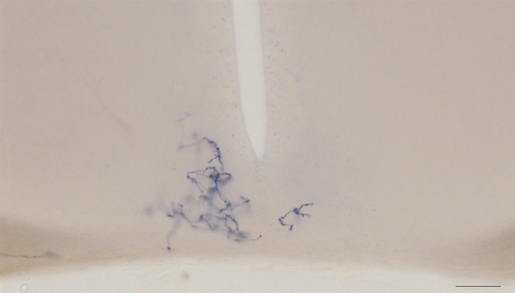
Figure 1. Representative image of a single ipRGC (dark blue fibers) that projects to the suprachiasmatic nucleus (SCN), the master clock in mammals.
Figure 2. Reconstruction of 20 ipRGC terminals in the SCN. Each color represents a single neuron, which can cover ~17% of the total volume of the SCN.
References
1. Shih-Kuo Chen, Tudor Constantin Badea, Samer Hattar. (2011). Photoentrainment and pupillary light reflex are mediated by distinct populations of ipRGCs. Nature. 476, 92-95. DOI: 10.1038/nature10206.
2. Diego Carlos Fernandez, Yi-Ting Chang, Samer Hattar, and Shih-Kuo Chen. (2016). Architecture of retinal projections to the central circadian pacemaker. Proceedings of the National Academy of Sciences of the United States of America (PNAS). 113(21), 6047-6052. DOI: 10.1073/pnas.1523629113.
Associate Professor Shih-Kuo Chen
Department of Life Science
alenskchen@ntu.edu.tw