In recent decades, ultrafast optical spectroscopy has provided a fundamental understanding of carrier dynamics (such as electrons and phonons) in many emergent materials, such as nanomaterials and 2D materials. Nevertheless, these materials typically possess spatial inhomogeneity, and it is therefore highly desirable to integrate ultrafast spectroscopy and optical microscopy to provide full spatiotemporal characterization of nanosized features. Here, we demonstrated a novel confocal pump-probe microscopy with a backscattering scheme for measuring isolated single nanomaterials. In contrast to the widely used transmission pump-probe microscopy, the confocal scheme provided axial sectioning capability, which enabled a subfemtoliter detection volume when combined with a tightly focusing objective.
However, the marriage between microscopy and spectroscopy is not a trivial task. For example, in a typical ultrafast spectroscopy setup, a pump and probe beams are employed, and nonlinear optical generation has typically been utilized to spectrally separate the pump/probe beams. Nevertheless, in a microscopy setup, when the wavelengths of the pump/probe beams are far from each other, the chromatic aberration of a high-NA microscopic objective lens becomes a serious issue.
In our study, to circumvent the aforementioned issues, we used sharp spectral filters to separate the spectra from the same optical pulses to create a pump beam and a probe beam. The pump and probe spectra and the original optical pulses (no filter) are shown in Figure 1(a). Because their spectra were directly divided from the full bandwidth of the original pulses, the wavelengths of the pump and probe beams were very similar, and the aberration effect was minimized. This method also reduced the complexity of the pump-probe microscopy geometry, eliminating the need, for example, for additional optical parametric oscillators (amplifiers).
For example, we studied the phonon dynamics of single gold nanoparticles by using a Ti:sapphire oscillator. Figure 1(b) shows the structure of the samples. Gold nanoparticles 80 nm in diameter were dispersed on a glass slide and coated with a PMMA film. As shown in Figure 1(b), the refractive indexes of the materials through which light penetrates were closely matched to each other except for the gold nanoparticles. Therefore, interface reflections were minimized, and the backscattering light in the confocal geometry was dominated by the gold nanoparticles. After pinpointing a single gold nanoparticle with the subfemtoliter spatial precision of confocal microscopy, the ultrafast temporal dynamics of the nanoparticle were determined by the pump-and-probe technique, i.e., collinearly aligning the pump and probe beams at the center of the gold nanoparticle and recording the scattering changes of the probe pulses (DS/S) as a function of the time delay to the pump pulses, as shown in Figure 1(c).
The physical process is as follows: 1) the strong pump pulses excite electrons in the gold nanoparticle; 2) the transient thermal energy initiates acoustic vibrations; and 3) the vibrations subsequently affect the scattering of the probe beam. The black and blue lines show the experimental and fitting curves, respectively. According to the fitting results, the oscillation frequency is 15.5 GHz with a damping time of 200 ps. With the knowledge that the transverse acoustic velocity of gold is 1200 m/s, the actual size of this gold nanoparticle is calculated as 77 nm.
Please note that the optical modulation signal (DS/S~10-3) in confocal pump-probe backscattering traces was orders of magnitude larger than in typical transmission-type pump-probe traces (DTr/Tr ~ , where Tr is the total intensity of the transmitted probe at the detector). This high sensitivity enabled, for the first time, the additional observation of signals due to the temperature evolution of the surrounding media, i.e., after single gold nanoparticles were heated by the pump beam, the energy flowed into the surrounding media. This phenomenon is shown by the existence of background relaxation in Figure 1(c) with a time constant of 384 ps, which is typical for thermal diffusion. Data fitting quantitatively shows a 2 K temperature increase and subsequent decrease in the surrounding media, providing new information on nanoscale thermal interactions. Our technique would pave a route for investigating the phonon dynamics of various nanomaterials.
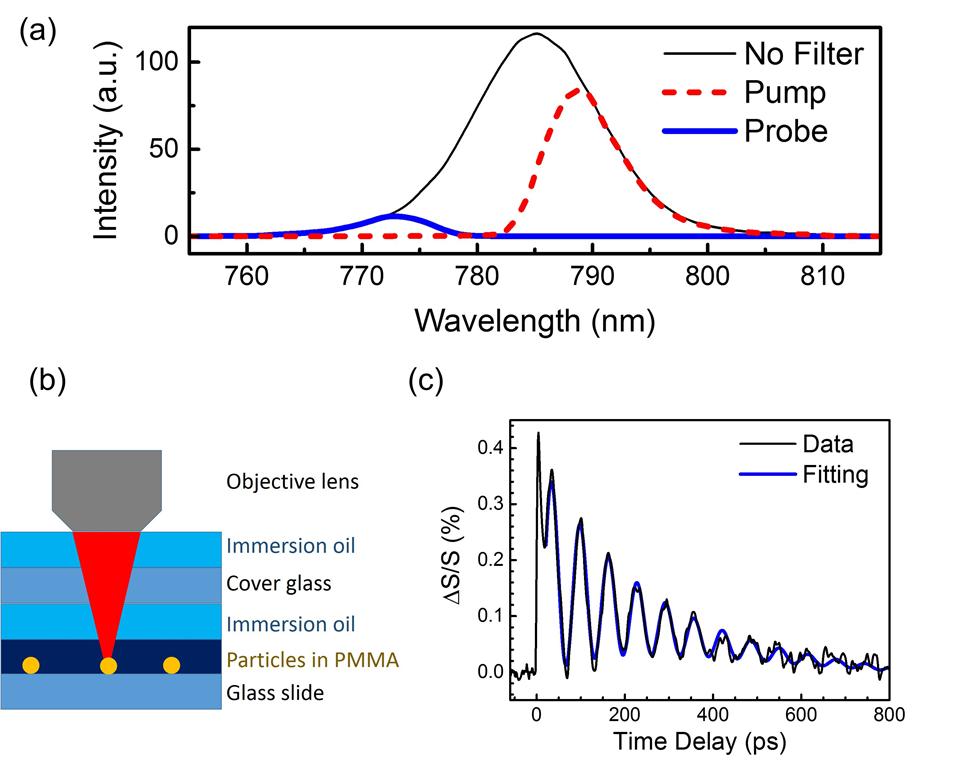
Figure 1 (a) The spectra of the optical pulses with no filter, with a long-wave-pass filter (for the pump), and with a short-wave-pass filter (for the probe). (b) A schematic of the sample structure with gold nanoparticles embedded in PMMA film. (c) A typical pump-probe trace of a gold single nanoparticle.
Reference
Kung-Hsuan Lin, Hao-Yu Cheng, Chi-Yuan Yang, Hung-Wei Li, Chih-Wei Chang, and Shi-Wei Chu. (2018). Phonon dynamics of single nanoparticles studied using confocal pump-probe backscattering. Applied Physics Letters, 113(17), 171906. DOI: 10.1063/1.5048669.
Dr. Kung-Hsuan Lin
Institute of Physics, Academia Sinica
Shi-Wei Chu
Professor, Department of Physics, National Taiwan University